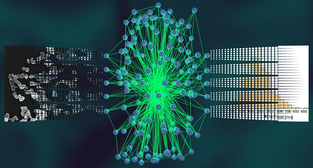
Schematic representation of a neural network. The inputs are EM images, the outputs are descriptors obtained from automated particle analysis.
Source: BAM, division Biophotonics
Even though it might seem trivial for a human, recognizing individual objects in front of a changing, non-uniform background is a challenging task for a computer. So far scientific images such as electron microscopy micrographs of nanoparticles typically have to be analyzed manually in a tedious process. An operator has to spend several hours outlining and measuring hundreds or even thousands of individual, small objects in these images by hand. This process is not only slow and tiring, but also prone to operator bias, which could skew the final results. Artificial neural networks, a form of machine learning/artificial intelligence that is for example also used in computer vision tasks required for self-driving cars, can learn and carry out these tasks as well with relatively high efficiency and accuracy. Once trained, such an artificial neural network can provide a fully automated, unbiased, and reproducible analysis of hundreds or even thousands of particles. However, the training process generally also requires a large amount of annotated data, which has to be prepared manually by humans again. In this publication, we present a workflow we developed for obtaining the fully trained artificial neural networks and the subsequent image analyses with minimal human interaction (approximately 10-15 minutes). For validation, we compare the performance of the neural network with the data collected through conventional means by humans from the same images. The presented methods should also be applicable to the analysis of other imaging techniques in the future.
Workflow towards automated segmentation of agglomerated, non‑spherical particles from electron microscopy images using artificial neural networks
Bastian Rühle, Julian Frederic Krumrey, Vasile-Dan Hodoroaba
published in Scientific Reports, Vol. 11, Issue 1, page 4942, 2021
BAM division Biophotonics, division Surface Analysis and Interfacial Chemistry


