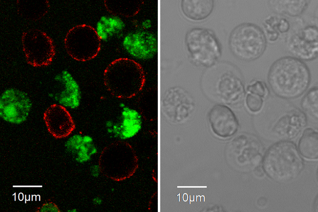
Left: Confocal Laser Scanning Microscopy images of living, specifically fluorophore-labelled hybridoma cells. Overlay of different fluorescence channels; red: living hybridoma cells expressing cell-surface IgG; green: cell produces antibodies that are selective for the hapten. Right: The same hybridoma cells in a light microscope.
Source: BAM, Department Analytical Chemistry; Reference Materials
Antibodies are large proteins, which are secreted to fight so-called antigens in the body of mammals in a polyclonal immune response. Due to their high specificity, antibodies are employed in highly selective (immuno-)analytical methods, medical diagnostics, and in therapeutic applications. For these purposes, monoclonal antibodies (mAbs) are preferred which stem from a single cell line and are created via the so-called hybridoma technique. Hybridoma cell screening and subcloning processes are of the most critical and time-consuming steps during production of monoclonal antibodies and the number of clones that can be tested is limited. For the development of our antibody-based analytical techniques, especially for the analysis of small molecules (haptens or incomplete antigens), we are devoted to improving and speeding up methods that will allow us – and other researchers – having a more straightforward access to hybridoma cells that produce the desired, highly selective, monoclonal antibodies.
At BAM, a research team headed by Dr. Rudolf Schneider demonstrated, that a sophisticated labelling technique for hybridoma cells combined with fluorescence-activated cell sorting (FACS) provides a powerful high-throughput method for the hapten-specific isolation of mAb-producing hybridoma cells. The two-step staining approach combines hapten-specific labelling (with a hapten−horseradish peroxidase conjugate subsequently labelled with a fluorophore-coupled anti-peroxidase antibody) and staining of those cells by a fluorophore-coupled anti-mouse IgG antibody that signals expression of membrane-bound antibodies (of IgG type). The labelled cells are easily distinguished by Confocal Laser Scanning Microscopy (CLSM) (see Figure). Using the FACS technique in a flow cytometer, hundreds of hybridoma cells producing hapten-specific mAbs can be rapidly sorted out from a cell mix and deposited as single-cell clones in wells of microtiter plates for further culture.
This high-throughput technique allows significant improvements in the generation of mAbs including increased isolation rate of antibody-producing hybridoma clones, guaranteed monoclonality of sorted cells, and reduced development times.
Hapten-Specific Single-Cell Selection of Hybridoma Clones by Fluorescence-Activated Cell Sorting for the Generation of Monoclonal Antibodies
Martin Dippong, Peter Carl, C. Lenz, J. A. Schenk, Katrin Hoffmann, Timm Schwaar, Rudolf J. Schneider, Maren Kuhne
Analytical Chemistry, 2017, 2017, 89 (7), pp 4007–4012
BAM Department 1 Analytical Chemistry; Reference Materials


